
Oligodendrocyte
| Oligodendrocyte | |
|---|---|
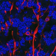
 Oligodendrocytes form the electrical insulation around the axons of CNS nerve cells. | |
| Details | |
| Location | Central nervous system |
| Identifiers | |
| Latin | oligodendrocytus |
| MeSH | D009836 |
| TH | H2.00.06.2.00003, H2.00.06.2.01018 |
| FMA | 83665 54540, 83665 |
| Anatomical terms of microanatomy | |
Oligodendrocytes (from Greek 'cells with a few branches'), also known as oligodendroglia, are a type of neuroglia whose main functions are to provide support and insulation to axons within the central nervous system (CNS) of jawed vertebrates. Their function is similar to that of Schwann cells, which perform the same task in the peripheral nervous system (PNS). Oligodendrocytes accomplish this by forming the myelin sheath around axons.[1] Unlike Schwann cells, a single oligodendrocyte can extend its processes to cover around 50 axons,[2] with each axon being wrapped in approximately 1 μm of myelin sheath. Furthermore, an oligodendrocyte can provide myelin segments for multiple adjacent axons.[1]
Oligodendrocytes are exclusively found in the CNS, which comprises the brain and spinal cord. Initially, it was originally thought that these cells were produced in the ventral neural tube, the embryonic precursor to the CNS. However, recent research suggests that oligodendrocytes originate from the ventral ventricular zone of the embryonic spinal cord, with some potential concentrations in the forebrain.[3] Notably, oligodendrocytes are the last type of cell to be generated in the CNS.[4] Oligodendrocytes were discovered by Pío del Río Hortega.[5][6]
Classification
[edit]Oligodendrocytes are a type of glial cell. They arise during development from oligodendrocyte precursor cells (OPCs),[7] which can be identified by their expression of a number of antigens, including the ganglioside GD3,[8][9][10] the NG2 chondroitin sulfate proteoglycan, and the platelet-derived growth factor-alpha receptor subunit (PDGF-alphaR).[11] Mature oligodendrocytes are broadly classified into either myelinating or non-myelinating satellite oligodendrocytes. Precursors and both mature types are typically identified by their expression of the transcription factor OLIG2.[12]
Development
[edit]Most oligodendrocytes develop during embryogenesis and early postnatal life from restricted periventricular germinal regions.[13] Oligodendrocyte formation in the adult brain is associated with glial-restricted progenitor cells, known as oligodendrocyte progenitor cells (OPCs).[14] Subventricular zone OPCs are activated and then migrate away from germinal[14] zones to populate both developing white and gray matter, where they differentiate and mature into myelin-forming oligodendrocytes.[10][15] However, it is not clear whether all oligodendrocyte progenitors undergo this sequence of events.[16]
Between midgestation and term birth in human cerebral white matter, three successive stages of the classic human oligodendrocyte lineage are found: OPCs, immature oligodendrocytes (non-myelinating), and mature oligodendrocytes (myelinating).[17] It has been suggested that some undergo apoptosis[18] and others fail to differentiate into mature oligodendrocytes but persist as adult OPCs.[19] Remarkably, oligodendrocyte population originated in the subventricular zone can be dramatically expanded by administering epidermal growth factor (EGF).[20][21]
Function
[edit]Myelination
[edit]
Mammalian nervous systems depend crucially on myelin sheaths, which reduce ion leakage and decrease the capacitance of the cell membrane, for rapid signal conduction.[22] Myelin also increases impulse speed, as saltatory propagation of action potentials occurs at the nodes of Ranvier in between Schwann cells (of the PNS) and oligodendrocytes (of the CNS). Furthermore, impulse speed of myelinated axons increases linearly with the axon diameter, whereas the impulse speed of unmyelinated cells increases only with the square root of the diameter. The insulation must be proportional to the diameter of the fibre inside. The optimal ratio of axon diameter divided by the total fiber diameter (which includes the myelin) is 0.6.[23]
Myelination is only prevalent in a few brain regions at birth and continues into adulthood. The entire process is not complete until about 25–30 years of age.[23] Myelination is an important component of intelligence, and white matter quantity may be positively correlated with IQ test results in children.[23] Rats that were raised in an enriched environment, which is known to increase cognitive flexibility, had more myelination in their corpus callosi.[24]
Immune function
[edit]Oligodendrocytes, best known for their role in myelinating axons in the central nervous system, also have important functions in immune regulation.[25] These cells can influence the immune environment by secreting cytokines and chemokines, which modulate the activity of various immune cells. Oligodendrocytes express receptors that allow them to respond to inflammatory signals, thereby participating in the brain's defense mechanisms. Additionally, they play a role in maintaining the blood-brain barrier and can contribute to the resolution of inflammation, highlighting their multifaceted role in both neural maintenance and immune responses.[25][26] While most research has focused on the immune functions of OPCs,[26][25] it is believed that oligodendrocytes themselves still possess significant immune functions.[25]
Metabolic support
[edit]Oligodendrocytes interact closely with nerve cells and provide trophic support by the production of glial cell line-derived neurotrophic factor (GDNF), brain-derived neurotrophic factor (BDNF), or insulin-like growth factor-1 (IGF-1).[27] They may also directly provide metabolites to neurons, as described by the lactate shuttle hypothesis.[28][29][30]
It is hypothesized that satellite oligodendrocytes (or perineuronal oligodendrocytes) are functionally distinct from other oligodendrocytes. They are not attached to neurons via myelin sheaths and, therefore, do not contribute to insulation. They remain opposed to neurons and regulate the extracellular fluid.[2] Satellite oligodendrocytes are considered to be a part of the grey matter whereas myelinating oligodendrocytes are a part of the white matter. They may support neuronal metabolism. Satellite oligodendrocytes may be recruited to produce new myelin after a demyelinating injury.[31]
Clinical significance
[edit]Diseases that result in injury to oligodendrocytes include demyelinating diseases such as multiple sclerosis and various leukodystrophies. Trauma to the body, e.g. spinal cord injury, can also cause demyelination. The immature oligodendrocytes, which increase in number during mid-gestation, are more vulnerable to hypoxic injury and are involved in periventricular leukomalacia.[32] This largely congenital condition of damage to the newly forming brain can therefore lead to cerebral palsy. In cerebral palsy, spinal cord injury, stroke and possibly multiple sclerosis, oligodendrocytes are thought to be damaged by excessive release of the neurotransmitter, glutamate.[33] Damage has also been shown to be mediated by N-methyl-D-aspartate receptors.[33] Oligodendrocyte dysfunction may also be implicated in the pathophysiology of schizophrenia and bipolar disorder.[34]
Oligodendrocytes are also susceptible to infection by the JC virus, which causes progressive multifocal leukoencephalopathy (PML), a condition that specifically affects white matter, typically in immunocompromised patients. Tumors of oligodendrocytes are called oligodendrogliomas. The chemotherapy agent Fluorouracil (5-FU) causes damage to the oligodendrocytes in mice, leading to both acute central nervous system (CNS) damage and progressively worsening delayed degeneration of the CNS.[35] [36] DNA methylation may also have a role in the degeneration of oligodendrocytes.[37]
Damage to myelin has been shown to exacerbate amyloid plaque accumulation, potentially placing age-related myelin decline as an upstream risk factor in Alzheimer's disease.[38] Oligodendrocytes also abundantly express components of the amyloidogenic pathway,[39][40] produce amyloid beta (Aβ), and contribute to plaque burden[40][41], which is relevant when considering therapeutic interventions for Alzheimer's disease.
See also
[edit]- 2',3'-Cyclic-nucleotide 3'-phosphodiesterase (CNPase)
- List of distinct cell types in the adult human body
- List of human cell types derived from the germ layers
- Schwann cells
- White matter
References
[edit]- ^ a b Carlson N (2010). Physiology of Behavior. Boston, MA: Allyn & Bacon. pp. 38–39. ISBN 978-0-205-66627-0.
- ^ a b Baumann N, Pham-Dinh D (April 2001). "Biology of oligodendrocyte and myelin in the mammalian central nervous system". Physiological Reviews. 81 (2): 871–927. doi:10.1152/physrev.2001.81.2.871. PMID 11274346.
- ^ Richardson WD, Kessaris N, Pringle N (January 2006). "Oligodendrocyte wars". Nature Reviews. Neuroscience. 7 (1): 11–18. doi:10.1038/nrn1826. PMC 6328010. PMID 16371946.
- ^ Thomas JL, Spassky N, Perez Villegas EM, Olivier C, Cobos I, Goujet-Zalc C, et al. (February 2000). "Spatiotemporal development of oligodendrocytes in the embryonic brain". Journal of Neuroscience Research. 59 (4): 471–476. doi:10.1002/(SICI)1097-4547(20000215)59:4<471::AID-JNR1>3.0.CO;2-3. PMID 10679785. S2CID 42473842.
- ^ Pérez-Cerdá F, Sánchez-Gómez MV, Matute C (2015). "Pío del Río Hortega and the discovery of the oligodendrocytes". Frontiers in Neuroanatomy. 9: 92. doi:10.3389/fnana.2015.00092. PMC 4493393. PMID 26217196.
- ^ James OG, Mehta AR, Behari M, Chandran S (June 2021). "Centenary of the oligodendrocyte". The Lancet. Neurology. 20 (6): 422. doi:10.1016/S1474-4422(21)00136-8. PMC 7610932. PMID 34022167.
- ^ Cameron-Curry P, Le Douarin NM (December 1995). "Oligodendrocyte precursors originate from both the dorsal and the ventral parts of the spinal cord". Neuron. 15 (6): 1299–1310. doi:10.1016/0896-6273(95)90009-8. PMID 8845154.
- ^ Curtis R, Cohen J, Fok-Seang J, Hanley MR, Gregson NA, Reynolds R, et al. (February 1988). "Development of macroglial cells in rat cerebellum. I. Use of antibodies to follow early in vivo development and migration of oligodendrocytes". Journal of Neurocytology. 17 (1): 43–54. doi:10.1007/BF01735376. PMID 3047324.
- ^ LeVine SM, Goldman JE (November 1988). "Ultrastructural characteristics of GD3 ganglioside-positive immature glia in rat forebrain white matter". The Journal of Comparative Neurology. 277 (3): 456–464. doi:10.1002/cne.902770310. PMID 3198802.
- ^ a b Hardy R, Reynolds R (April 1991). "Proliferation and differentiation potential of rat forebrain oligodendroglial progenitors both in vitro and in vivo". Development (Cambridge, England). 111 (4): 1061–1080. doi:10.1242/dev.111.4.1061. PMID 1879350.
- ^ Pringle NP, Mudhar HS, Collarini EJ, Richardson WD (June 1992). "PDGF receptors in the rat CNS: during late neurogenesis, PDGF alpha-receptor expression appears to be restricted to glial cells of the oligodendrocyte lineage". Development. 115 (2): 535–551. doi:10.1242/dev.115.2.535. PMID 1425339.
- ^ Yokoo H, Nobusawa S, Takebayashi H, Ikenaka K, Isoda K, Kamiya M, et al. (May 2004). "Anti-human Olig2 antibody as a useful immunohistochemical marker of normal oligodendrocytes and gliomas". The American Journal of Pathology. 164 (5): 1717–1725. doi:10.1016/S0002-9440(10)63730-3. PMC 1615653. PMID 15111318.
- ^ Vallstedt A, Klos JM, Ericson J (January 2005). "Multiple dorsoventral origins of oligodendrocyte generation in the spinal cord and hindbrain". Neuron. 1. 45 (1): 55–67. doi:10.1016/j.neuron.2004.12.026. hdl:10616/40454. PMID 15629702. S2CID 7971750.
- ^ a b Menn B, Garcia-Verdugo JM, Yaschine C, Gonzalez-Perez O, Rowitch D, Alvarez-Buylla A (July 2006). "Origin of oligodendrocytes in the subventricular zone of the adult brain". The Journal of Neuroscience. 26 (30): 7907–7918. doi:10.1523/JNEUROSCI.1299-06.2006. PMC 6674207. PMID 16870736.
- ^ Levison SW, Goldman JE (February 1993). "Both oligodendrocytes and astrocytes develop from progenitors in the subventricular zone of postnatal rat forebrain". Neuron. 10 (2): 201–12. doi:10.1016/0896-6273(93)90311-e. PMID 8439409.
- ^ Franklin RJ, Ffrench-Constant C (November 2008). "Remyelination in the CNS: from biology to therapy". Nature Reviews. Neuroscience. 9 (11): 839–855. doi:10.1038/nrn2480. PMID 18931697.
- ^ Barateiro A, Fernandes A (September 2014). "Temporal oligodendrocyte lineage progression: in vitro models of proliferation, differentiation and myelination". Biochimica et Biophysica Acta (BBA) - Molecular Cell Research. 1843 (9): 1917–1929. doi:10.1016/j.bbamcr.2014.04.018. PMID 24768715.
- ^ Barres BA, Hart IK, Coles HS, Burne JF, Voyvodic JT, Richardson WD, et al. (November 1992). "Cell death in the oligodendrocyte lineage". Journal of Neurobiology. 23 (9): 1221–1230. doi:10.1002/neu.480230912. PMID 1469385.
- ^ Wren D, Wolswijk G, Noble M (January 1992). "In vitro analysis of the origin and maintenance of O-2Aadult progenitor cells". The Journal of Cell Biology. 116 (1): 167–76. doi:10.1083/jcb.116.1.167. PMC 2289266. PMID 1730741.
- ^ Gonzalez-Perez O, Romero-Rodriguez R, Soriano-Navarro M, Garcia-Verdugo JM, Alvarez-Buylla A (August 2009). "Epidermal growth factor induces the progeny of subventricular zone type B cells to migrate and differentiate into oligodendrocytes". Stem Cells. 27 (8): 2032–2043. doi:10.1002/stem.119. PMC 3346259. PMID 19544429.
- ^ Gonzalez-Perez O, Alvarez-Buylla A (June 2011). "Oligodendrogenesis in the subventricular zone and the role of epidermal growth factor". Brain Research Reviews. 67 (1–2): 147–156. doi:10.1016/j.brainresrev.2011.01.001. PMC 3109119. PMID 21236296.
- ^ Sokol S. "The Physiology and Pathophysiology of Multiple Sclerosis". Multiple Sclerosis: Physiological Tutorial. Archived from the original on 2012-07-16. Retrieved 2012-04-29.
- ^ a b c Fields RD (March 2008). "White matter matters". Scientific American. 298 (3): 42–49. Bibcode:2008SciAm.298c..54D. doi:10.1038/scientificamerican0308-54. PMID 18357821.
- ^ Juraska JM, Kopcik JR (May 1988). "Sex and environmental influences on the size and ultrastructure of the rat corpus callosum". Brain Research. 450 (1–2): 1–8. doi:10.1016/0006-8993(88)91538-7. PMID 3401704. S2CID 2720782.
- ^ a b c d Zeis T, Enz L, Schaeren-Wiemers N (June 2016). "The immunomodulatory oligodendrocyte". Brain Research. Evolution of Myelin. 1641 (Pt A): 139–148. doi:10.1016/j.brainres.2015.09.021. PMID 26423932.
- ^ a b Zveik O, Rechtman A, Ganz T, Vaknin-Dembinsky A (July 2024). "The interplay of inflammation and remyelination: rethinking MS treatment with a focus on oligodendrocyte progenitor cells". Molecular Neurodegeneration. 19 (1): 53. doi:10.1186/s13024-024-00742-8. PMC 11245841. PMID 38997755.
- ^ Bradl M, Lassmann H (January 2010). "Oligodendrocytes: biology and pathology". Acta Neuropathologica. 119 (1): 37–53. doi:10.1007/s00401-009-0601-5. PMC 2799635. PMID 19847447.
...oligodendrocytes can provide trophic support for neurons by the production of glial cell line-derived neurotrophic factor (GDNF), brain-derived neurotrophic factor (BDNF), or insulin-like growth factor-1 (IGF-1).
- ^ Philips T, Rothstein JD (September 2017). "Oligodendroglia: metabolic supporters of neurons". The Journal of Clinical Investigation. 127 (9): 3271–3280. doi:10.1172/JCI90610. PMC 5669561. PMID 28862639.
- ^ Fünfschilling U, Supplie LM, Mahad D, Boretius S, Saab AS, Edgar J, et al. (April 2012). "Glycolytic oligodendrocytes maintain myelin and long-term axonal integrity". Nature. 485 (7399): 517–521. Bibcode:2012Natur.485..517F. doi:10.1038/nature11007. PMC 3613737. PMID 22622581.
- ^ Nave KA, Asadollahi E, Sasmita A (December 2023). "Expanding the function of oligodendrocytes to brain energy metabolism". Current Opinion in Neurobiology. 83: 102782. doi:10.1016/j.conb.2023.102782. PMID 37703600.
- ^ Szuchet S, Nielsen JA, Lovas G, Domowicz MS, de Velasco JM, Maric D, et al. (December 2011). "The genetic signature of perineuronal oligodendrocytes reveals their unique phenotype". The European Journal of Neuroscience. 34 (12): 1906–1922. doi:10.1111/j.1460-9568.2011.07922.x. PMC 4286392. PMID 22132705.
- ^ Kinney HC, Back SA (September 1998). "Human oligodendroglial development: relationship to periventricular leukomalacia". Seminars in Pediatric Neurology. 5 (3): 180–189. doi:10.1016/s1071-9091(98)80033-8. PMID 9777676.
- ^ a b Káradóttir R, Attwell D (April 2007). "Neurotransmitter receptors in the life and death of oligodendrocytes". Neuroscience. 145 (4): 1426–1438. doi:10.1016/j.neuroscience.2006.08.070. PMC 2173944. PMID 17049173.
- ^ Tkachev D, Mimmack ML, Ryan MM, Wayland M, Freeman T, Jones PB, et al. (September 2003). "Oligodendrocyte dysfunction in schizophrenia and bipolar disorder". Lancet. 362 (9386): 798–805. doi:10.1016/S0140-6736(03)14289-4. PMID 13678875. S2CID 7511585.
- ^ "Chemotherapy-induced Damage to the CNS as a Precursor Cell Disease" Archived 2011-12-27 at the Wayback Machine by Dr. Mark D. Noble, University of Rochester
- ^ Han R, Yang YM, Dietrich J, Luebke A, Mayer-Pröschel M, Noble M (2008). "Systemic 5-fluorouracil treatment causes a syndrome of delayed myelin destruction in the central nervous system". Journal of Biology. 7 (4): 12. doi:10.1186/jbiol69. PMC 2397490. PMID 18430259.
- ^ Fodder K, de Silva R, Warner TT, Bettencourt C (June 2023). "The contribution of DNA methylation to the (dys)function of oligodendroglia in neurodegeneration". Acta Neuropathologica Communications. 11 (1): 106. doi:10.1186/s40478-023-01607-9. PMC 10311741. PMID 37386505.
- ^ Depp C, Sun T, Sasmita AO, Spieth L, Berghoff SA, Nazarenko T, et al. (June 2023). "Myelin dysfunction drives amyloid-β deposition in models of Alzheimer's disease". Nature. 618 (7964): 349–357. Bibcode:2023Natur.618..349D. doi:10.1038/s41586-023-06120-6. PMC 10247380. PMID 37258678.
- ^ Gazestani V, Kamath T, Nadaf NM, Dougalis A, Burris SJ, Rooney B, et al. (September 2023). "Early Alzheimer's disease pathology in human cortex involves transient cell states". Cell. 186 (20): 4438–4453.e23. doi:10.1016/j.cell.2023.08.005. PMC 11107481. PMID 37774681.
- ^ a b Sasmita AO, Depp C, Nazarenko T, Sun T, Siems SB, Ong EC, et al. (August 2024). "Oligodendrocytes produce amyloid-β and contribute to plaque formation alongside neurons in Alzheimer's disease model mice". Nature Neuroscience: 1–7. doi:10.1038/s41593-024-01730-3. PMID 39103558.
- ^ Rajani RM, Ellingford R, Hellmuth M, Harris SS, Taso OS, Graykowski D, et al. (July 2024). "Selective suppression of oligodendrocyte-derived amyloid beta rescues neuronal dysfunction in Alzheimer's disease". PLOS Biology. 22 (7): e3002727. doi:10.1371/journal.pbio.3002727. PMC 11265669. PMID 39042667.
Further reading
[edit]- Raine CS (1991). "Oligodendrocytes and central nervous system myelin.". In Davis RL, Robertson DM (eds.). Textbook of Neuropathology (second ed.). Baltimore, Maryland: Williams and Wilkins. pp. 115–141.
External links
[edit]- The Department of Neuroscience at Wikiversity
- NIF Search – Oligodendrocyte Archived 2013-07-03 at the Wayback Machine via the Neuroscience Information Framework